Research Projects

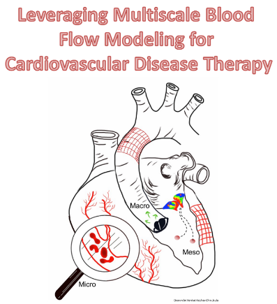
Our primary research area is cardiovascular biomechanics with a focus on multiscale blood flow modeling. Leveraging my strong interdisciplinary computational expertise with complementary in vitro investigations, I seek to develop hemodynamics-driven, evidence-based mechanistic methodologies to understand, detect, diagnose and treat cardiovascular disease. I envision a future where personalized medicine will deliver on its promise and I plan to contribute towards it by incorporating synergistic and interdisciplinary technologies that improve patient outcomes.

In the MCFL, we focus on understanding blood flow (hemodynamics) under healthy and pathological conditions to understand, detect, diagnose and treat cardiovascular disease. We collaborate very closely with clinicians (cardiology, cardiac surgery, vascular surgery, neurosurgery) to ensure our research is always translational, i.e. patient-focused.

We use a combination of tools such as computational fluid dynamics (CFD) modeling of blood flow, virtual surgery and optimization, reduced order modeling of the cardiovascular system, predictive modeling, 3D printing and rapid prototyping.
Leveraging Multiscale blood Flow Modeling for Cardiovascular Disease TherapySome of the current / previous projects in the lab focus on:

  • Heart failure therapy
  • Life-support devices (Extracorporeal Membrane Oxygenation or ECMO)
  • Cerebral aneurysms
  • Abdominal aortic aneurysms
  • Platelet activation / thrombosis
  • Medical devices / implants
  • Congenital heart diseases
  • Virtual surgery and patient management
  • Blood particulate interactions
  • 3D printing and rapid prototyping of cardiovascular blood vessel models

Looking for graduate / undergraduate researchers!

Potential projects include (but not limited to): heart failure, cardiogenic shock, stroke, atherosclerosis, atrial fibrillation, 3D computational fluid dynamics simulations of blood flow in the heart and various blood vessels, reduced order modeling of the cardiovascular network, Lagrangian tracking of individual red blood cells / platelets, platelet activation modeling, development of computational techniques, machine learning, 3d printing, rapid prototyping.

Preferred background

Curiosity about the cardiovascular system and how medical therapy works, experience with computational analysis software such as MATLAB / Python / C / C++, design software such as Solidworks / Autocad, computational modeling (such as CFD or finite element modeling and meshing), 3D printing, rapid prototyping. Familiarity with fluid mechanics, cardiovascular physiology, cardiovascular pathologies and/or medical therapies is ideal. Inquisitiveness and an open mind are essential!

If you are interested, please email me at vchivukula@fit.edu with your resume and transcripts

Our Projects


3D Reconstruction of Cerebral Aneurysms from 2D Angiographic Imaging using Deep Learning

Ruth White and Marcello Mattei


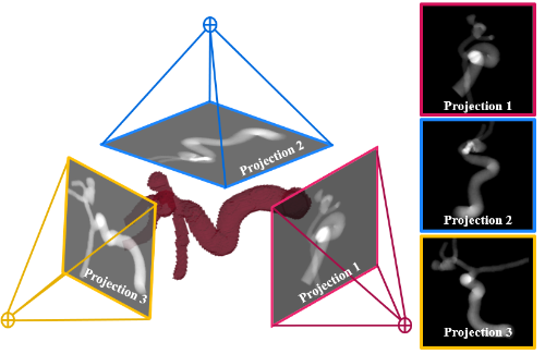
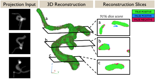
Clinicians rely on 2D imaging techniques to lead and guide endovascular procedures. Cerebral vasculature is highly 3 dimensional with complex curvature and tortuosity; however, imaging techniques “flatten” the complex vasculature. Thus, our goal is to reconstruct vasculature in 3D from a sparse number of 2D angiographic images. 

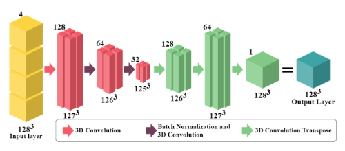
To do this, we have created a vast database of simulated angiographic images from 159 unique patient-specific aneurysms. We then created a custom AI to successfully reconstruct the aneurysm and surrounding vasculature. This same network can be applied to any part of the cardiovascular system, including coronary arteries, aortas, left ventricles, and peripheral vasculature. 

The 3D reconstructed vasculature and 3D angiography can then be correlated with hemodynamic information such as velocity, vorticity, and shear stress. 


American Heart Association - Wikipedia 

Funded by AHA Predoctoral Fellowship 24PRE1242524

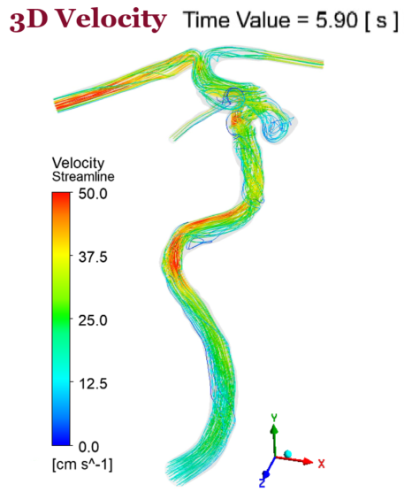
Investigating Cerebral Aneurysm Hemodynamics

Ruth White and Erin Smith


Brain aneurysms are the ballooning of a blood vessel in the brain. This can be dangerous because there is a chance the aneurysm could rupture or cause a clot which could lead to brain damage or even death. Using specialized tools, doctors have methods to treat the aneurysms from the inside of the blood vessels. To observe these aneurysms and determine treatment strategies, doctors use angiographic images, which show them the size and shape of the aneurysm; however, these images don't provide much information about how the blood flows inside the aneurysm. This blood flow information is important for treatment because it could let us know about the chance of rupture. 
 
We use computers to model the blood flow inside of aneurysms which gives us a deeper understanding of how the blood flows in these aneurysms. We will first look at a large range of patient aneurysms, angiographic images, and their blood flow. We can then relate these images to identify patterns and trends in the blood flow within the aneurysm. However, this takes too much time (days or even weeks) to check for every patient, so we will use deep learning (another form of artificial intelligence, AI) to speed up this process to make it a useful treatment tool. 


See our SPIE Medical Imaging 2023 Proceedings Paper and Presentation

See our SPIE Medical Imaging 2022 Proceedings Paper and Presentation


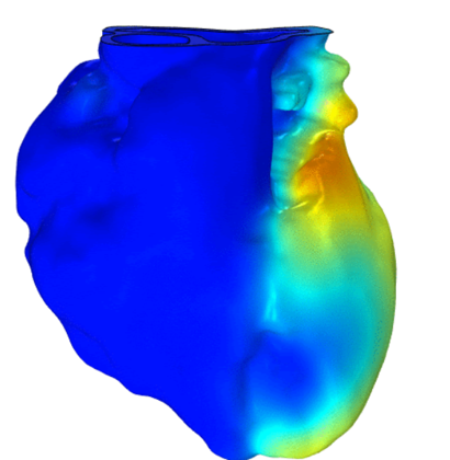
Modeling Cardiac Heat Transfer and Fluid Dynamics to Optimize Heart Transplantation

Juan Rodriguez Paez, Kaitlyn Dunn, Lasya Gopagani, Ruth White


Despite advancements in preservation technology, many donor hearts remain unused in part due to a lack of suitable preservation strategies for transportation, yet the temperature of a heart during organ transplantation is mostly unknown. I am performing computational fluid dynamics (CFD) and heat transfer simulations to optimize the temperature for organs during their transplant process. The first step of this research has been focused on the heart in reported patient-specific cases with the current transplant techniques. These simulations allow us to understand how different hearts might behave under these physical and environmental conditions. Also, the results obtained from the simulations allow us to predict the biological implications for the organs undergoing those specific processes.


Exploring Left Ventricular Assist Device (LVAD) Patient Hemodynamic Interplay

Jasmine Martinez and Holly Grant


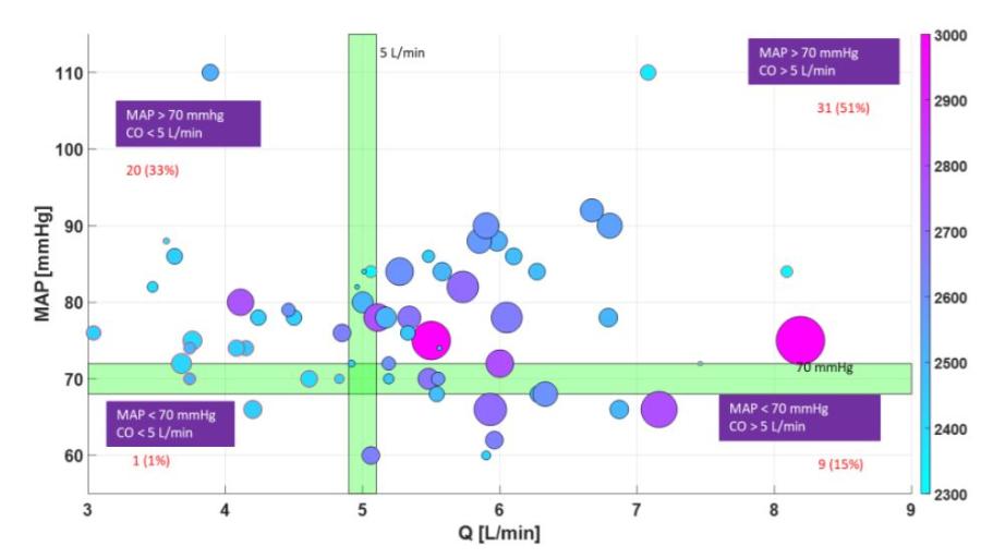
Our research involves helping patients who suffer from stage D heart failure and are aided by a left ventricular assist device (LVAD). We have an in-house developed model which allows us to mimic patient-specific conditions. Using this model, we are able to explore components that play a role in the function of optimizing the LVAD to improve the patient’s hemodynamics. 

See our ASAIO Journal Publication here! 


Geometric Deep Learning for Optimizing Left Ventricular Assist Devices (LVAD) Therapy for Heart Failure

Rachel Hillner, Marcello Vittorio Mattei Di Eugenio, Ben Diaz 


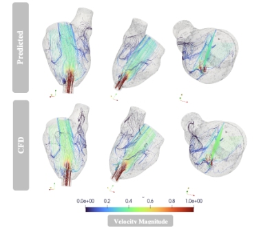
 Computational Fluid Dynamics, CFD, is a popular tool used in biomedical engineering to better understand how blood traverses different geometries within the human body. For instance, it can provide great insight to how abnormalities and surgical implants impact blood flow, i.e. blood flow in aneurysms and in a LVAD (left ventricular assist device) implanted LV (left ventricle). However, CFD can be quite time consuming and computational expensive, thus making patient specific investigations difficult. We are looking to train a neural network to predict blood flow through a variety of geometries, thus minimizing computational expense and making patient specific analysis feasible in a clinical setting.  

This project is funded by an American Heart Associatin (AHA) Career Development Award #937847 to Dr. Chivukula 

 American Heart Association - Wikipedia


Assessing Performance the Pediatric Heart Assist Device, EXCOR 

Natalie Hill and Rachel Hillner


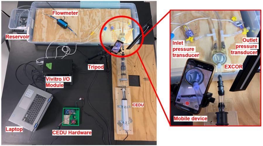
Our research involves studying the EXCOR, the only FDA-approved pediatric heart assist device. We are using a ViVitro Cardiovascular Superpump to test the EXCOR under different hemodynamic situations so we can better understand what causes the EXCOR to malfunction.  We then developed an app that can detect these abnormalities in the EXCOR. From this data and our app, clinicians will be able to identify these situations before the patient is severely affected by a malfunctioning EXCOR.


Improving Blockage Detection in the Heart-Lung Machine

Ruth White


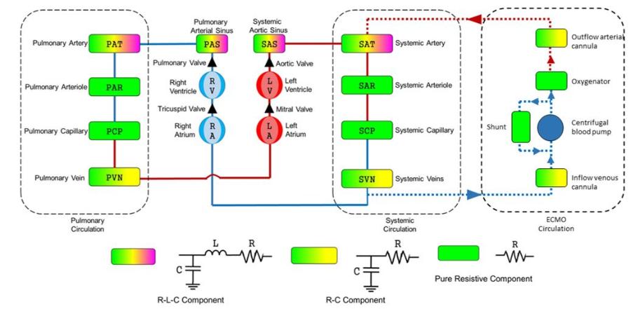
Heart-lung machines (ECMO) provide cardiac and respiratory support to patients whose heart or lungs are inadequate for blood flow or gas exchange. The current methods for monitoring system blockages have limitations. We are exploring the idea that the flow in a small shunt tube in the ECMO circuit will be affected by oxygenator clotting or other ECMO circuit changes. The goal of our study is to test the hypothesis that blood flow rates in the shunt could be used to monitor for circuit obstructions using a computational model of the patient-ECMO system.

Modeling the Cardiovascular-Renal System Interaction for Implantable Artificial Kidney Therapy


The renal system plays important roles in the homeostasis of the body and is a significant component of the cardiovascular system. Receiving roughly 20% of the cardiac output under normal conditions, it has its own feedback mechanisms to regulate blood flow and pressure within it. Our research focuses on modeling the dynamic response of the renal system to optimize design and interaction of an Implantable Artificial Kidney Device for renal disease therapy. 

Funded by a NIH NIDDK R01 in collaboration with Vanderbilt University and the University of California San Francisco

Visualizing CFD Flow Patterns in App

Eshan Vipuil

An important aspect of hemodynamic analysis is visualizing flow patterns in a range of scenarios. This project aims to develop an accessible and free to use webapp where clinicians and researchers can visualize flow from CFD.